UK-first ultra-high definition CT scanner will be used on mummies
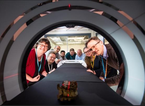
THE first ultra-high definition CT scanner of its kind in the UK has been installed at the University of Bradford School of Archaeological and Forensic Sciences, allowing scientists to examine centuries-old artefacts and remains in greater detail than ever before.
There are only around 50 FujiFilm NewTom 7G Plus Cone Beam CT machines in the world, and the University’s is the first to be installed in the UK and the first to be used outside of healthcare.
The cutting-edge technology means archaeological and forensic scientists can scan the most fragile human remains and even the most delicate of artefacts to see what’s hidden inside, without causing any damage.
Project Lead Professor Andrew Wilson, Chair of Archaeology and Forensic Sciences, said: “The possibilities in terms of imaging are far-reaching, not just for human remains such as bone with pathological lesions, but also instances where soft tissue survives including with historic potted specimens and mummies and of course with archaeological artefacts and forensic evidential materials."
The new capability will provide one of the first dedicated facilities in the UK for archaeologists and heritage scientists to routinely undertake cross-sectional imaging of wide-ranging materials. It is the only currently available Cone Beam CT instrument that allows merged and stitched capture.
Example artefacts used so far include fused archaeological vertebrae and a sealed 19th century box from a recent, local dig, which revealed a pair of glasses inside, with pristine glass lenses and tortoiseshell frames.
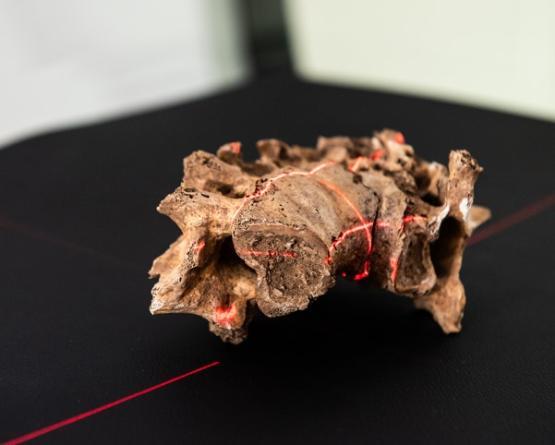
Pictured above: Fused archaeological vertebrae from the collections of the Biological Anthropology Research Centre exhibiting pathological lesions that were featured in the landmark Digitised Diseases resource.
Dr Cathy Batt, Head of Archaeological and Forensic Sciences, said: “The glasses were in a box inside protective packaging but we can’t look directly inside the box because it’s so fragile.
“What this unit does is allow us to look at archaeological and forensic materials in a lot more detail.
“For example, we can study cremation urns where ash and soil is often mixed with human remains and we will be able to determine the different materials, and whether pyre goods were incorporated.”
The instrumentation is part of £3 million worth of investment that the University has received through CapCo, the Capability for Collections fund, part of the Arts and Humanities Research Council’s (AHRC) allocation of world class laboratories funding.
It’s not unusual for archaeologists to use such technology but they mostly have to rely on using healthcare scanners out of clinic hours when they are not needed for patients.
Robert Janaway, Associate Professor in Archaeological Sciences said: “Using healthcare scanners isn’t ideal. It’s not only morally problematic given the current backlog of cases in the NHS but in practical terms, the radiation strength used in patient-fronted facilities is much lower, in order to protect the patient.
“Having our own unit means that we can modify the radiation exposure to see evidence more clearly.”
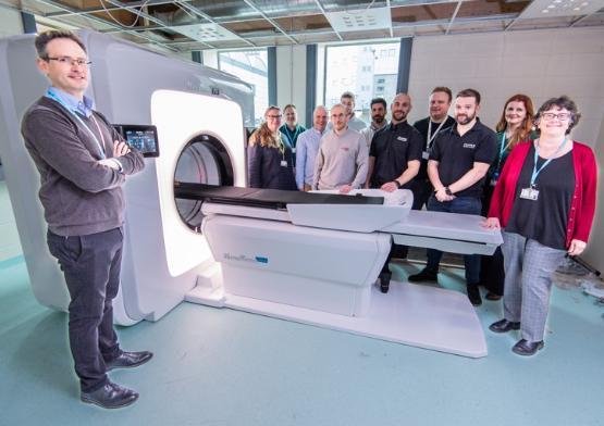
Pictured above: Professor Andrew Wilson with academics from Archaeological Sciences and representatives from FujiFilm
The scanner will serve as a capability to allow academics and students to further develop outstanding work in archaeological techniques and technology, something the University has already been recognised for with the Queen’s Anniversary Prize.
Dr Batt said: “We see the Queen’s Anniversary Prize as a springboard, rather than an end point. Because this is the first scanner of its kind in the UK, students will be able to learn at the very edge of advances in technology.
“There will be uses for this that we haven’t yet thought of and as a non-destructive testing method it will almost certainly lead us to groundbreaking discoveries.”
Bradford has a national and internationally recognised capability for Human Bioarchaeology and Digital Collections and this complements our work in Visualising Heritage, meaning we can collaborate with other institutions and commercial companies so that they can access our facilities for their research and knowledge exchange, further adding to the University’s reputation as a world leader.“
The instrument is sited in a prominent position on campus – within the University Analytical Centre and adjacent to the Norcroft Conferencing Suite.
Dr Richard Telford, Director of the University Analytical Centre, said: "We're delighted to be hosting the Cone Beam CT scanner within the Analytical Centre. This will sit alongside existing infrastructure that includes other instruments funded through the CapCo investment. It will strengthen the cross-disciplinary research focus of the facility which serves as a hub for Analytical Science at the University."
However, getting such a piece of world-class equipment onto campus was no mean feat.
Mark Howroyd, project manager at Fujifilm, said: "To install a Cone Beam CT scanner in five days is impressively quick and a credit to both the University and to our engineering team. There is a lot of planning involved when installing a machine like this, from the layout of the room, right down to cable routes, radiation protection and ergonomics of use. Working with the University has been a very positive experience, everyone has been really helpful and cooperative. It is a delight to see the research team so engaged and eager to use the Cone Beam CT"
Stuart McTavish, Team Leader Modality Applications at Fujifilm, said: "We are building a strong partnership with the University of Bradford. We're excited to be involved in helping bring this piece of cutting-edge technology to the University."